
![]()
In December 2019 the city of Wuhan (China) became the center of corona virus and with the time goes, today entire world is affected by this COVID-19. At the same time availability of limited COVID-19 testing kits, we need to rely on other diagnosis measures. Since COVID-19 attacks the epithelial cells that line our respiratory tract, we can use X-rays to analyze the health of a patient’s lungs. Also nearly all hospitals have X-ray imaging machines, it could be possible to use X-rays to test for COVID-19 without the dedicated test kits.
In this case, AI and image processing becomes useful tools in the detection of the disease. However application of such tools at premature stage does not provide a confidence in diagnosis but it helps us to prioritize a test to be performed and reduction in the data-set on which laboratory test need to perform. Recently, CT and Chest X-rays can play an important role in the diagnosis of COVID-19.
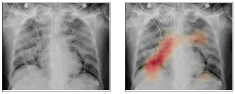
Figure 1 shows manual findings of an abnormality in the chest x-ray. However this manual feature extraction is very time consuming as well as resource hungry (i.e domain export manual resource). Hence manual extraction does not work at this stage. In this case AI/ML and image processing technology helps a lot to make process automatic and less resource hungry (i.e domain export manual resource).
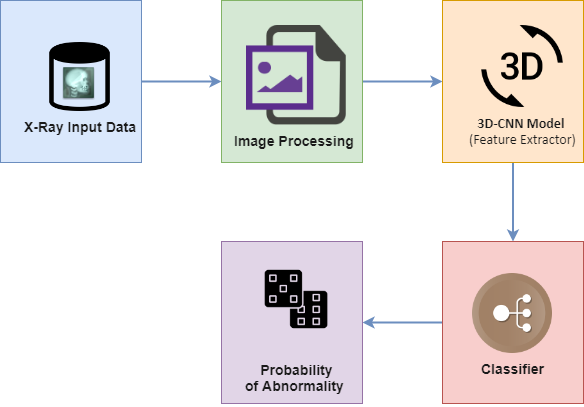
In order to speed up the discovery of disease mechanisms, Firstly Chest X-Ray Classifier can be used to detect abnormalities then after extract textural features of the altered lung parenchima and finally combine both findings to figure out specific signatures of the Covid-19 virus.
How could abnormality to be detected in X-ray images?
In this blog we will cover a first part of pipeline chest x-ray classification. In any deep learning-based classifier design. First stage is to prepare the database. For abnormality classification we need both abnormal and normal data. Abnormal (covid-19) infected chest X-ray images are collected from COVID-19 image data [1] collected by Paul, Morrison and Dao and put them into “./Dataset/covid/” folder. Chest X-ray images of healthy persons are collected from kaggle pneumonia data [2]. From that we have taken x-ray samples of healthy persons and put them into “./Dataset/normal/” folder.
Following is the broad level structure of algorithm.
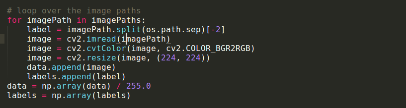
To make a dataset trainable, we take image from folder and resize it into 224 x 224 x 3 and generate it’s corresponding one hot labeling from the mane of folder.
As we have less amount of label data available one of the nest way is to fine tune the existing deep learning model rather than develop and train a new model from scratch. Therefore, we initialize our VGGNet model and add our own classifier layers on top of it.
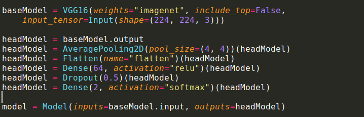
Finally, we divide our sample data into 80% train and 20% test and fine tune the model for binary classification. The prepared model is not so accurate as the less data available but the performance of the model can definitely increase by increasing amount of available data.
Disclaimer: The above section does not claim, nor does it intend to “solve”, COVID-19 detection. The purpose if the blog is to encourage the Deep learning and computer vision researcher about putting their effort in solving the real time problem and help a medical domain. The purpose of the above section is to develop a helper which can help to prioritize a test to be performed and reduction in the data-set on which laboratory test need to perform. However, the algorithm can be put into actual use once proper following of TRIPOD guidelines [3] and proper False positive and false negative analysis. Because in this case false negative is very harmful as compared to false positive (the intention is not to justify false positive).
References:
[1] https://github.com/ieee8023/covid-chestxray-dataset
[2] https://www.kaggle.com/paultimothymooney/chest-xray-pneumonia
[3] https://www.equator-network.org/reporting-guidelines/tripod-statement/